Trusted Worldwide Questions & Answers
ARDMS AB-Abdomen Dumps - Pass the Abdomen Sonography Examination in 2026
The ARDMS AB-Abdomen exam, officially the Abdomen Sonography Examination, is part of the Registered Diagnostic Medical Sonographer certification path. It is designed for candidates who want to demonstrate knowledge and clinical understanding in abdominal sonography. This exam matters because it validates the skills needed to support accurate imaging, interpretation, and patient care in abdominal ultrasound practice.
For sonography professionals and candidates preparing for ARDMS certification, a focused study plan can make a major difference. Understanding the exam structure and topic areas helps you build confidence and improve your readiness before test day.
| # | Exam Topics | Sub-Topics | Approximate Weightage (%) |
|---|---|---|---|
| 1 | Anatomy, Perfusion, and Function | Abdominal organ anatomy, Normal blood flow and perfusion, Organ function relationships | 35% |
| 2 | Pathology, Vascular Abnormalities, Trauma, and Postoperative Anatomy | Common abdominal pathology, Vascular abnormalities, Trauma findings, Postoperative anatomy | 30% |
| 3 | Abdominal Physics | Ultrasound principles, Image optimization, Artifacts and interpretation | 20% |
| 4 | Clinical Care, Practice, and Quality Assurance | Patient care, Scanning practice, Quality assurance and safety | 15% |
The exam tests both knowledge and practical judgment across abdominal sonography. Candidates must understand anatomy, recognize abnormalities, apply ultrasound physics, and follow proper clinical and quality assurance practices. Strong exam performance depends on both content mastery and the ability to apply that knowledge in realistic testing situations.
How QA4Exam.com Helps You Pass
QA4Exam.com offers an Exam PDF with actual questions and answers plus an Online Practice Test for the ARDMS AB-Abdomen exam. These resources help you study with a real exam simulation so you can become familiar with the question style and pacing. The content is designed to be up to date and includes verified answers to support accurate review. With timed practice, you can improve time management and build confidence before the actual test. Using both the PDF and practice test together can help you prepare more effectively and aim to pass on your first attempt.
Frequently Asked Questions
What is the ARDMS AB-Abdomen exam?
The ARDMS AB-Abdomen exam is the Abdomen Sonography Examination. It is part of the Registered Diagnostic Medical Sonographer certification and focuses on abdominal sonography knowledge and practice.
Who is this exam for?
This exam is for candidates pursuing the Registered Diagnostic Medical Sonographer certification who want to demonstrate competence in abdomen sonography.
Is the ARDMS AB-Abdomen exam difficult?
The exam can be challenging because it covers anatomy, pathology, physics, and clinical practice. Candidates who study the core topics and practice with exam-style questions are better prepared for the level of difficulty.
Can I pass with only braindumps?
Braindumps alone are not the best approach. You should also understand the concepts behind the questions so you can handle different question formats and apply the knowledge correctly on exam day.
Do I need hands-on experience to prepare well?
Hands-on experience is very helpful because it strengthens your understanding of anatomy, scanning practice, and clinical care. Even if you are using dumps and practice tests, real-world exposure can improve your confidence and accuracy.
Are QA4Exam.com dumps and practice tests enough to prepare?
QA4Exam.com materials are useful for focused preparation, especially when you want actual questions and answers plus a practice test format. For best results, combine them with topic review so you understand the exam areas in depth.
How do the QA4Exam.com files help with passing on the first attempt?
The Exam PDF and Online Practice Test help you review verified answers, practice under time pressure, and get familiar with the exam style. This combination supports better readiness and can improve your chances of passing on the first attempt.
What format do the QA4Exam.com materials use?
QA4Exam.com provides an Exam PDF with questions and answers and an Online Practice Test that simulates the exam experience. This gives you both study convenience and interactive practice.
The questions for AB-Abdomen were last updated on Jul 21, 2026.
- Viewing page 1 out of 33 pages.
- Viewing questions 1-5 out of 165 questions
Which clinical indication is most consistent with the finding depicted in this image?
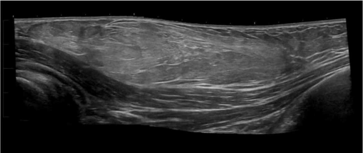
The ultrasound image shows disruption of the normal fibrillar echotexture of a muscle or tendon, consistent with a soft tissue injury such as a muscle or tendon tear. There is likely hypoechoic fluid consistent with a hematoma or edema, which commonly results from blunt or direct trauma.
This image is typical of a traumatic injury (e.g., partial or complete tendon rupture or muscle strain/tear). These findings are frequently encountered in athletic injuries or blunt force trauma and correlate strongly with the clinical history of trauma.
Key sonographic features suggestive of trauma:
Discontinuity or heterogeneity of normal striated muscle or tendon pattern
Hypoechoic or anechoic area representing hematoma or fluid collection
Retraction of muscle or tendon ends (in full-thickness tears)
Surrounding soft tissue edema
Differentiation from other options:
B . Focal pain: While pain may be a symptom, trauma is the more definitive and primary clinical indication for the findings shown.
C . Palpable abnormality: May suggest a mass or cystic lesion (e.g., lipoma, abscess), not typically the appearance shown here.
D . Decreased range of motion: May be present secondarily, but not the most consistent or primary clinical indication in this case.
Bianchi S, Martinoli C. Ultrasound of the Musculoskeletal System. Springer, 2007. Chapters on Muscle and Tendon Injuries.
American Institute of Ultrasound in Medicine (AIUM) Practice Parameter for the Performance of a Musculoskeletal Ultrasound Examination, 2020.
Radiopaedia.org. Muscle tear (ultrasound): https://radiopaedia.org/articles/muscle-tear-ultrasound
Where in the neck are most thyroid cancer recurrences found?
Most thyroid cancer recurrences are found in the ipsilateral neck---particularly in the central (level VI) or lateral (levels II-V) compartments on the same side as the original malignancy.
According to AIUM Practice Parameters:
''Post-thyroidectomy recurrence most frequently occurs ipsilateral to the original tumor, commonly involving regional lymph nodes.''
AIUM Practice Parameter for Thyroid and Neck Ultrasound, 2020.
American Thyroid Association (ATA) Guidelines for Thyroid Cancer Management, 2015.
Which condition is most likely associated with a common bile duct measuring 5 mm?
A common bile duct (CBD) measuring up to 5 mm is considered normal in most patients under age 60. Some references allow for up to 6 mm, especially post-cholecystectomy or in older individuals. Significant dilation (suggestive of obstruction) typically exceeds these measurements.
According to Rumack's Diagnostic Ultrasound:
''The normal common bile duct measures up to 5--6 mm, with slight increases considered normal after cholecystectomy or with advancing age.''
Rumack CM, Wilson SR, Charboneau JW, Levine D. Diagnostic Ultrasound. 5th ed. Elsevier, 2017.
AIUM Practice Parameter for Abdominal Ultrasound, 2020.
Which vascular condition is most likely associated with the sonographic findings demonstrated in this image?
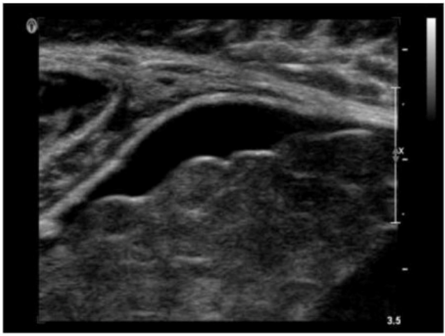
The ultrasound image demonstrates a tubular, anechoic structure coursing anterior to the left portal vein and heading toward the anterior abdominal wall. This is consistent with a recanalized umbilical vein, which is an important collateral pathway that reopens in cases of portal hypertension.
Normally, the umbilical vein becomes obliterated after birth and forms the ligamentum teres. However, in the setting of significant portal hypertension, the umbilical vein may recanalize and serve as a collateral route to decompress the portal system.
Sonographic features of a recanalized umbilical vein:
Anechoic, tubular structure in the ligamentum teres fissure
Seen anterior to the left portal vein
Color Doppler confirms hepatofugal venous flow
Associated with signs of portal hypertension (e.g., splenomegaly, varices)
Differentiation from other options:
A . Budd-Chiari syndrome: Involves hepatic vein outflow obstruction; ultrasound shows absent or narrowed hepatic veins and may have caudate lobe hypertrophy.
B . Splenic artery aneurysm: Typically visualized near the splenic hilum as a pulsatile cystic mass; Doppler shows arterial flow.
D . Median arcuate ligament syndrome: Involves compression of the celiac axis; best assessed with Doppler showing elevated velocities on expiration.
Rumack CM, Wilson SR, Charboneau JW, Levine D. Diagnostic Ultrasound. 5th Edition. Elsevier, 2018. Chapter: Portal Hypertension and Collaterals, pp. 101--104.
American Institute of Ultrasound in Medicine (AIUM). Practice Parameter for the Performance of a Vascular Ultrasound Examination, 2020.
Radiopaedia.org. Recanalized umbilical vein: https://radiopaedia.org/articles/recanalised-umbilical-vein
Which pancreatic condition is commonly associated with complete or partial atresia of the duodenum?
Annular pancreas is a congenital anomaly in which pancreatic tissue encircles the second part of the duodenum, potentially causing partial or complete duodenal obstruction (atresia). It is due to abnormal migration of the ventral pancreatic bud.
According to Rumack's Diagnostic Ultrasound:
''Annular pancreas results from failure of the ventral pancreatic bud to rotate properly, leading to encirclement of the duodenum.''
Rumack CM, Wilson SR, Charboneau JW, Levine D. Diagnostic Ultrasound. 5th ed. Elsevier, 2017.
Moore KL, Clinically Oriented Anatomy. 8th ed. Wolters Kluwer, 2018.
---
Unlock All Questions for ARDMS AB-Abdomen Exam
Full Exam Access, Actual Exam Questions, Validated Answers, Anytime Anywhere, No Download Limits, No Practice Limits
Get All 165 Questions & Answers