Trusted Worldwide Questions & Answers
ARDMS AE-Adult-Echocardiography Dumps - Pass AE Adult Echocardiography Examination in 2026
The ARDMS AE-Adult-Echocardiography - AE Adult Echocardiography Examination is part of the Registered Diagnostic Cardiac Sonographer certification path. It is designed for candidates who want to demonstrate strong knowledge of adult echocardiography concepts, clinical application, and sonographic interpretation. Passing this exam supports professional credibility and helps validate your readiness to work in cardiac ultrasound settings. For many candidates, it is an important step toward advancing in diagnostic cardiac sonography.
Exam Topics and Approximate Weightage
| # | Exam Topics | Sub-Topics | Approximate Weightage (%) |
|---|---|---|---|
| 1 | Anatomy and Physiology | Cardiac chambers and valves; blood flow and circulation; cardiac cycle basics | 24% |
| 2 | Pathology | Common adult cardiac disease processes; structural abnormalities; functional impact on echo findings | 22% |
| 3 | Clinical Care and Safety | Patient preparation; infection control; safety considerations and professional practice | 16% |
| 4 | Measurement Techniques, Maneuvers, and Sonographic Views | Standard echocardiographic views; measurement methods; imaging maneuvers and acquisition techniques | 24% |
| 5 | Instrumentation, Optimization, and Contrast | Machine controls and image optimization; transducer and system settings; contrast-related concepts | 14% |
This exam tests both theoretical knowledge and practical understanding of adult echocardiography. Candidates must know cardiac anatomy, recognize pathology, apply safe clinical practices, and understand how to obtain and optimize diagnostic images. It also measures your ability to use measurement techniques and sonographic views correctly in real exam scenarios.
How QA4Exam.com Helps You Pass
QA4Exam.com provides an Exam PDF with actual questions and answers plus an Online Practice Test built to help you prepare for the ARDMS AE-Adult-Echocardiography exam efficiently. The practice test gives you a real exam simulation so you can get familiar with the question style and pacing before test day. You also get up-to-date questions, verified answers, and a focused way to review the exam areas that matter most. By practicing in a timed format, you can improve time management and reduce exam-day stress. These tools are designed to help you study smarter and aim for first-attempt success.
Frequently Asked Questions
What is the ARDMS AE Adult Echocardiography Examination?
It is the AE Adult Echocardiography Examination under the ARDMS Registered Diagnostic Cardiac Sonographer certification path. It assesses adult echocardiography knowledge, imaging understanding, and clinical application.
Who is this exam for?
It is for candidates pursuing the Registered Diagnostic Cardiac Sonographer certification and those who want to prove competence in adult echocardiography.
Is the AE-Adult-Echocardiography exam difficult?
It can be challenging because it covers anatomy, pathology, safety, measurement techniques, and instrumentation. Strong preparation and practice with exam-style questions can make it much more manageable.
Can I pass with only braindumps?
Braindumps alone are not the best approach. You should use them as a study aid along with understanding the topics, reviewing explanations, and practicing with realistic exam questions.
Do I need hands-on experience to prepare well?
Hands-on experience is very helpful because this exam includes practical echocardiography concepts. Even if you are studying from materials, real-world familiarity improves understanding and recall.
Are QA4Exam.com dumps and practice tests enough to prepare?
They are highly useful for targeted review, question practice, and exam simulation, but combining them with broader study can improve confidence and readiness even more.
How do QA4Exam.com dumps and the online practice test help with first-attempt success?
They help you study with actual questions and answers, verify your knowledge, and practice under timed conditions. That combination supports better retention and stronger time management on exam day.
What format do the QA4Exam.com products come in?
QA4Exam.com offers an Exam PDF and an Online Practice Test. These formats are designed to make review flexible, convenient, and close to the real exam experience.
The questions for AE-Adult-Echocardiography were last updated on Jul 20, 2026.
- Viewing page 1 out of 28 pages.
- Viewing questions 1-5 out of 139 questions
Which type of rendering is primarily utilized with three-dimensional echocardiography?
Comprehensive and Detailed Explanation From Exact Extract:
Three-dimensional echocardiography (3D echo) primarily uses volume rendering to provide a realistic and spatially accurate representation of cardiac structures. Volume rendering processes a full dataset of voxels (3D pixels) to produce detailed images, allowing clinicians to visualize complex anatomical relationships in real time.
Surface rendering is another technique but primarily used in post-processing to create a solid surface model; it is less used in live 3D echocardiography.
Planar and external rendering are not standard terms in 3D echocardiography.
This information is presented in the 'Textbook of Clinical Echocardiography, 6e', Chapter on Advanced Echocardiographic Imaging Techniques20:400-405Textbook of Clinical Echocardiography.
Which method is useful for obtaining a good quality pulmonary vein spectral Doppler waveform for evaluation of diastolic function?
Comprehensive and Detailed Explanation From Exact Extract:
Pulmonary vein Doppler signals have low velocity and low frequency components that can be filtered out by standard Doppler wall filters. To obtain a good quality spectral Doppler waveform for diastolic function evaluation, the wall filter settings should be lowered or adjusted to allow low frequency signals to be detected and displayed clearly.
Non-imaging transducers and continuous wave Doppler are not appropriate for pulmonary vein Doppler because spatial resolution and site localization are required. Filtering out low frequency signals would degrade the quality of the pulmonary vein waveform.
This is detailed in ASE Doppler imaging and diastolic function assessment protocols12:ASE Diastolic Function Guidelinesp.85-9016:Textbook of Clinical Echocardiography, 6ep.125-130.
The respirometer should be turned on when assessing which possible disease process(es)?
A respirometer monitors the respiratory cycle and is essential when evaluating diseases in which respiratory variation affects echocardiographic measurements, such as pericardial effusion and cardiac tamponade. In tamponade, respiratory changes in mitral and tricuspid inflows, as well as variations in inferior vena cava size, are key diagnostic features.
Congestive heart failure, ischemic cardiomyopathy, and mitral valve diseases do not require synchronization with respiration for diagnosis or quantification and are not reliant on respirometer use.
This recommendation is outlined in ASE pericardial disease guidelines and echocardiography procedural protocols16:Textbook of Clinical Echocardiography, 6ep.280-28512:ASE Pericardial Disease Guidelinesp.300-305.
Which finding is shown in this image?
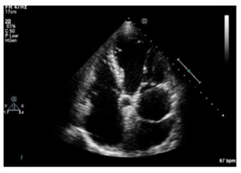
The echocardiographic image shows a mobile, highly echogenic, mesh-like structure within the right atrium consistent with the Chiari network. The Chiari network is an embryologic remnant of the right valve of the sinus venosus, appearing as a fenestrated, reticulated membrane that is usually thin and mobile, found near the orifice of the inferior vena cava or the coronary sinus.
This structure is benign and often an incidental finding but can be confused with thrombus or atrial tumors. Unlike left atrial thrombus, which appears as a more solid, immobile mass often located in the left atrial appendage, the Chiari network is mobile and located in the right atrium. Cor triatriatum is a rare congenital membrane dividing the left atrium into two chambers and appears differently on echocardiography. Artifact refers to non-anatomic echoes which do not persist or move consistently.
Recognition of Chiari network is important to avoid misdiagnosis, and its characteristics are well described in echocardiography literature such as the 'Textbook of Clinical Echocardiography' and ASE imaging guidelines16:Textbook of Clinical Echocardiography, 6ep.400-40212:ASE Guidelines on Cardiac Massesp.150-155.
A. Sinus venosus atrial septal defect
The echocardiographic image shows a typical atrial septal defect located in the central portion of the atrial septum, best classified as a secundum atrial septal defect (ASD). Secundum ASDs are the most common type, occurring in the fossa ovalis region.
Sinus venosus ASDs are located near the superior vena cava or inferior vena cava junctions, coronary sinus ASDs involve unroofing of the coronary sinus, and primum ASDs occur low in the atrial septum near the atrioventricular valves.
These anatomic distinctions are critical for diagnosis and surgical planning and are detailed in adult congenital heart disease and echocardiography references16:Textbook of Clinical Echocardiography, 6ep.565-57012:ASE Adult Congenital Guidelinesp.400-410.
Unlock All Questions for ARDMS AE-Adult-Echocardiography Exam
Full Exam Access, Actual Exam Questions, Validated Answers, Anytime Anywhere, No Download Limits, No Practice Limits
Get All 139 Questions & Answers