Trusted Worldwide Questions & Answers
ARDMS SPI Dumps - Pass Sonography Principles and Instrumentation Exam in 2026
The ARDMS SPI - Sonography Principles and Instrumentation exam is part of the Registered Diagnostic Cardiac Sonographer certification path. It is designed for candidates who want to validate their knowledge of ultrasound principles, image quality, and instrumentation basics. This exam matters because it supports accurate, safe, and effective sonographic practice. Strong performance on SPI can help you move forward with greater confidence in your sonography career.
Exam Topics Overview
| # | Exam Topics | Sub-Topics | Approximate Weightage (%) |
|---|---|---|---|
| 1 | Perform Ultrasound Examinations | Patient setup and positioning, transducer selection, scanning workflow | 22% |
| 2 | Manage Ultrasound Transducers | Transducer types, handling and care, frequency selection | 18% |
| 3 | Optimize Sonographic Images | Gain and depth adjustments, focus and dynamic range, artifact recognition | 24% |
| 4 | Apply Doppler Concepts | Color Doppler basics, spectral Doppler interpretation, flow direction and velocity | 20% |
| 5 | Provide Clinical Safety & Quality Assurance | Bioeffects and safety, infection control, equipment quality checks | 16% |
This exam tests both theoretical knowledge and practical understanding of ultrasound principles. Candidates should be ready to interpret imaging concepts, manage equipment correctly, and apply safe clinical practices. It also checks whether you can make sound decisions about image optimization and Doppler use in real exam scenarios.
How QA4Exam.com Helps You Pass
QA4Exam.com offers an Exam PDF with actual questions and answers plus an Online Practice Test to help you prepare for the ARDMS SPI exam efficiently. The practice materials are designed to simulate the real exam experience so you can get familiar with the question style, pacing, and pressure. You also benefit from up-to-date questions and verified answers, which helps you focus on what matters most. By practicing with timed questions, you can improve time management and reduce surprises on exam day. This combination gives you a practical path to aim for a first-attempt pass.
Frequently Asked Questions
1. What is the ARDMS SPI exam?
The ARDMS Sonography Principles and Instrumentation exam measures core ultrasound principles, instrumentation knowledge, and image optimization skills for the Registered Diagnostic Cardiac Sonographer path.
2. Is the SPI exam difficult?
Many candidates find it challenging because it tests both concepts and applied knowledge. With focused preparation and regular practice, it becomes much more manageable.
3. Can I pass with only braindumps?
Braindumps alone are not the best approach. You should use them as part of a broader study plan that includes understanding the concepts and practicing exam-style questions.
4. Do I need hands-on experience to prepare for SPI?
Hands-on exposure can help you understand the material better, but the exam itself focuses on principles and instrumentation knowledge. Good study resources can still help you prepare effectively.
5. Are the QA4Exam.com dumps enough to pass on the first attempt?
The QA4Exam.com Exam PDF and Online Practice Test are strong preparation tools, especially when used consistently. They help you review real exam-style questions, verify answers, and build confidence for a first-attempt pass.
6. What format do the QA4Exam.com practice materials use?
QA4Exam.com provides an Exam PDF with questions and answers and an Online Practice Test that helps you simulate the exam environment and practice time management.
7. What if I do not pass on the first try?
If you need to retake the exam, reviewing weak areas and practicing with updated questions can help you improve before your next attempt.
The questions for SPI were last updated on Jul 20, 2026.
- Viewing page 1 out of 43 pages.
- Viewing questions 1-5 out of 215 questions
What determines the resonant frequency of a pulsed wave transducer?
The resonant frequency of a pulsed wave transducer is determined by the thickness of the piezoelectric element and the speed of sound within that element. The resonant frequency is inversely proportional to the element thickness and directly proportional to the speed of sound in the material. Thinner elements and higher sound speeds result in higher resonant frequencies, while thicker elements and lower sound speeds result in lower resonant frequencies. Reference:
ARDMS Sonography Principles and Instrumentation guidelines
Kremkau, F. W. (2015). Diagnostic Ultrasound: Principles and Instruments. Elsevier.
Which setting is the most likely cause of the artifact displayed in this image?
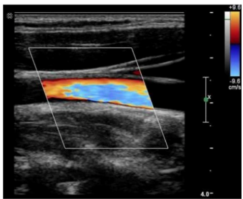
Comprehensive and Detailed Explanation From Exact Extract:
The image shows color aliasing, where the colors abruptly change within the vessel indicating wrap-around of Doppler shifts. This happens when the velocity scale (PRF) is set too low, causing velocities exceeding the Nyquist limit to alias.
According to sonography instrumentation reference:
''Aliasing occurs in color Doppler when the flow velocities exceed the Nyquist limit, commonly due to a low velocity scale (PRF).''
Therefore, the correct answer is D: Velocity scale set too low.
---
How is the wavelength affected when switching from a 10 MHz transducer to a 5 MHz transducer?
Comprehensive and Detailed Explanation From Exact Extract:
Wavelength is inversely proportional to frequency according to the equation:
Wavelength () = Propagation speed (c) / Frequency (f)
If frequency decreases from 10 MHz to 5 MHz, the wavelength increases by a factor of 2 (doubles).
Principles and Instrumentation state:
'As frequency decreases, wavelength increases. Halving the frequency results in doubling the wavelength.'
Therefore, the correct answer is D: Doubles.
---
Which adjustment will reduce the appearance of posterior shadowing artifact?
Comprehensive and Detailed Explanation From Exact Extract:
Spatial compounding uses multiple scan angles to create an image. By combining information from different angles, it can reduce shadowing artifacts caused by highly attenuating structures.
According to sonography instrumentation reference:
''Spatial compounding reduces artifacts such as posterior shadowing by averaging images acquired from multiple insonation angles.''
Therefore, the correct answer is A: Increasing spatial compounding.
---
What is the result of increasing the wall filter setting during Doppler sampling?
Comprehensive and Detailed Explanation From Exact Extract:
The wall filter in Doppler ultrasound is designed to eliminate low-frequency signals, typically associated with motion artifacts such as vessel wall or tissue motion. These low-frequency signals are not part of the desired blood flow signal and can interfere with accurate Doppler display.
When the wall filter setting is increased, it removes these low-frequency signals from the Doppler spectrum. However, increasing the wall filter too much can also eliminate true low-velocity flow information, leading to a loss of clinically relevant data.
This principle is described in official sonography Principles and Instrumentation references:
'Increasing the wall filter will reduce the display of low-frequency Doppler shifts, which are typically associated with slow-moving structures. These low-frequency signals can represent either slow blood flow or tissue motion artifacts.'
Therefore, the correct answer is D: Reduced display of low-frequency shifts.
Unlock All Questions for ARDMS SPI Exam
Full Exam Access, Actual Exam Questions, Validated Answers, Anytime Anywhere, No Download Limits, No Practice Limits
Get All 215 Questions & Answers